Tecnologias para o Diagnóstico da Radiodermite: uma Revisão Sistemática
Tecnologías para el diagnóstico de radiodermatitis: una revisión sistemática

Como Citar
Baixar Citação

Este trabalho está licenciado sob uma licença Creative Commons Attribution-NonCommercial-NoDerivatives 4.0 International License.
##submission.authorBio##
Artigos mais lidos pelo mesmo(s) autor(es)
- Rafael Chieza Fortes García, Rafael Melo de Oliveira, Eduardo Camilo Martínez, Eduardo Borba Neves, Precisão da equação de estimativa do VO₂ em adultos jovens , Archivos de Medicina (Manizales): v. 20 n. 1 (2020)
- Leandro de Lima e Silva, Eduardo Borba Neves, Luciano Alonso Valente dos Santos, Jurandir Baptista Da Silva, Rodrigo Gomes de Souza Vale, Rodolfo de Alkmim Moreira Nunes, The reproducibility of the 1hz gps in the aciclic displacement with intermittent intensity , Archivos de Medicina (Manizales): v. 21 n. 1 (2021): .
- Karielly Cássia de Almeida, Carmen Caroline Rasera, Wagner Luis Ripka, Tainá Ribas Mélo, Eduardo Borba Neves, RESPIRATORY FUNCTION IN CHILDREN WITH CEREBRAL PALSY , Archivos de Medicina (Manizales): v. 23 n. 1 (2023): .


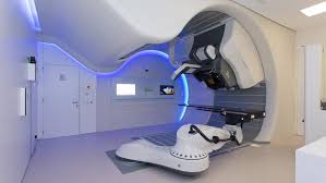
Objetivo: a radiodermite ou radiodermatite é uma reação celular inflamatória desencadeada pela radioterapia, que é uma das alternativas para tratamento de vários tipos de cânceres. O objetivo deste trabalho foi analisar os principais métodos de diagnóstico de radiodermite com uso de ferramentas tecnológicas, quanto a equipamentos utilizados, forma de análise clínica, parâmetros desenvolvidos e produto final. Materiais e métodos: optou-se pela revisão sistemática de caráter exploratório e abordagem qualitativa. A busca por estudos em periódicos internacionais foi realizada no mês de julho de 2019, nas bases de dados MEDLINE e SCOPUS. Foram utilizados os descritores “radiodermatitis” e “acute radiation dermatitis”, para a busca. Os critérios de inclusão foram: artigo, escrita em inglês, publicação nos últimos dez anos (01/2009 a 07/2019), e conteúdo relacionado ao diagnóstico de radiodermite causada por radioterapia de aplicação exclusiva. Resultados: esta revisão encontrou estudos sobre as seguintes ferramentas tecnológicas: avaliação citológica, avaliação de fluxo sanguíneo, machine learning, escala fotográfica, imagem térmica, dose de irradiação e espectrofotometria. O uso de imagens fotográficas e térmicas tem se consolidado como ferramenta de diagnóstico importante na área médica. Essas ferramentas tecnológicas têm se mostrado aplicáveis ao diagnóstico da radiodermite, com seu efeito potencializado por processamento digital de imagens e algoritmos de inteligência artificial. Conclusões: a análise aqui descrita demonstra a necessidade de desenvolvimento tecnológico para delineamento e padronização do processo de diagnóstico da radiodermite, o que permitiria tratamento precoce e manutenção do engajamento do indivíduo na terapia antineoplásica.
Visão geral 945 | Visualizações de PDF 73
Downloads
- Oselame GB, Sanches IJ, Kuntze A, Neves EB. Software for automatic diagnostic prediction of skin clinical images based on ABCD rule. Biosci J 2017; 33(4):1065-1078. DOI: 10.14393/BJ-v33n4a2017-34738
- Sekiguchi K, Akahane K, Ogita M, Haga C, Ito R, Arai S, et al. Efficacy of heparinoid moisturizer as a prophylactic agent for radiation dermatitis following radiotherapy after breast-conserving surgery: a randomized controlled trial. Jpn J Clin Oncol 2018; 48(5):450-457. DOI: 10.1093/jjco/hyy045
- Lee TF, Sung KC, Chao PJ, Huang YJ, Lan JH, Wu HY, et al. Relationships among patient characteristics, irradiation treatment planning parameters, and treatment toxicity of acute radiation dermatitis after breast hybrid intensity modulation radiation therapy. PloS one 2018; 13(7):e0200192. DOI: 10.1371/journal.pone.0200192
- Beamer LC, Grant M. Longitudinal trends in skin-related and global quality of life among women with breast radiodermatitis: a pilot study. Eur J Oncol Nurs 2018; 33:22-27. DOI: 10.1016/j.ejon.2018.01.008
- Lilla C, Ambrosone CB, Kropp S, Helmbold I, Schmezer P, von Fournier D, et al. Predictive factors for late normal tissue complications following radiotherapy for breast cancer. Breast Cancer Res Treat 2007; 106(1):143-150. DOI: 10.1007/s10549-006-9480-9
- Reddy J, Lindsay W, Berlind C, Ahern C, Smith B. Applying a Machine Learning Approach to Predict Acute Toxicities During Radiation for Breast Cancer Patients. Int J Radiat Oncol Biol Phys 2018; 102(3):S59. DOI: 10.1016/j.ijrobp.2018.06.167
- Whiting PF, Rutjes AW, Westwood ME, Mallett S, Deeks JJ, Reitsma JB, et al. QUADAS-2: a revised tool for the quality assessment of diagnostic accuracy studies. Ann Intern Med 2011;155(8):529-536. DOI: 10.7326/0003-4819-155-8-201110180-00009
- Maillot O, Leduc N, Atallah V, Escarmant P, Petit A, Belhomme S, et al. Evaluation of acute skin toxicity of breast radiotherapy using thermography: results of a prospective single-centre trial. Cancer Radiother 2018;22(3):205-210. DOI: 10.1016/j.canrad.2017.10.007
- Partl R, Jonko B, Schnidar S, Schöllhammer M, Bauer M, Singh S, et al. 128 SHADES OF RED: Objective Remote Assessment of Radiation Dermatitis by Augmented Digital Skin Imaging. Stud Health Technol Inform 2017; 236:363-374. DOI: 10.3233/978-1-61499-759-7-363
- Sun LM, Huang CJ, Chen HY, Chang GH, Tsao MJ. Evaluating the consistency of location of the most severe acute skin reaction and highest skin dose measured by thermoluminescent dosimeter during radiotherapy for breast cancer. Med Dos 2016; 41(3):216-220. DOI: 10.1016/j.meddos.2016.02.002
- González-Sanchis A, Brualla-González L, Sanchez-Carazo JL, Gordo-Partearroyo JC, Esteve-Martínez A,Vicedo-González A, et al. Evaluation of acute skin toxicity in breast radiotherapy with a new quantitative approach. Radiother Oncol 2017; 122(1):54-59. DOI: 10.1016/j.radonc.2016.09.019
- Huang CJ, Hou MF, Luo KH, Wei SY, Huang MY, Su SJ, et al. RTOG, CTCAE and WHO criteria for acute radiation dermatitis correlate with cutaneous blood flow measurements. Breast J 2015; 24(3):230-236. DOI: 10.1016/j.breast.2015.01.008
- Shumway D, Kapdia N, Do T, Griffith K, Feng M, Jagsi R, et al. Development of a photonumeric scale for acute radiation dermatitis in breast cancer patients. Int J Radiat Oncol Biol Phys 2014; 90(1):S238. DOI: 10.1016/j.ijrobp.2014.05.847
- Vano‐Galvan S, Fernandez‐Lizarbe E, Truchuelo M, Diaz‐Ley B, Grillo E, Sanchez V, et al. Dynamic skin changes of acute radiation dermatitis revealed by in vivo reflectance confocal microscopy. J Eur Acad Dermatol Venereol 2013;27(9):1143-1150. DOI: 10.1111/j.1468-3083.2012.04680.x
- Yoshikawa N, Inomata T, Shimbo T, Takahashi M, Uesugi Y, Juri H, et al. Appropriate evaluation of and risk factors for radiation dermatitis in breast cancer patients receiving hypofractionated whole-breast irradiation after breast-conserving surgery. Breast cancer. 2014; 21(2):170-176. DOI: 10.1007/s12282-012-0366-x
- Graham PH, Plant NA, Graham JL, Browne LH, Borg M, Capp A, et al. Digital photography as source documentation of skin toxicity: An analysis from the Trans Tasman Radiation Oncology Group (TROG) 04.01 Post‐Mastectomy Radiation Skin Care Trial. J Med Imaging Radiat Oncol 2012; 56(4):458-463. DOI: 10.1111/j.1754-9485.2012.02365.x
- Lucey P, Zouzias C, Franco L, Chennupati SK, Kalnicki S, McLellan BN. Practice patterns for the prophylaxis and treatment of acute radiation dermatitis in the United States. Support Care Cancer 2017; 25(9):2857-2862. DOI: 10.1007/s00520-017-3701-0
- Silva-De Andrade KB, Lima-Francz AC, Dos Santos-Grellmann M, Cortez-Belchior P, Araújo-De Oliveira J, Do Nascimento-Wassita D. Consulta de enfermagem: avaliação da adesão ao autocuidado dos pacientes submetidos à radioterapia. Rev enferm UERJ 2014;22(5):622-8. DOI: 10.12957/reuerj.2014.11227
- Capitani G, Sehnem E, Rosa C, Matos F, Reis VM, Neves EB. Osgood-schlatter Disease Diagnosis by Algometry and Infrared Thermography. Open Sports Sci J 2017; 10(2):223-228. DOI: 10.2174/1875399X01710010223
- Neves EB, Moreira TR, Lemos R, Vilaça-Alves J, Rosa C, Reis VM. Using skin temperature and muscle thickness to assess muscle response to strength training. Rev Bras Med Esporte 2015;21(5):350-4. DOI: 10.1590/1517-869220152105151293
- Mendes R, Sousa N, Almeida A, Vilaça-Alves J, Reis VM, Neves EB. Thermography: a technique for assessing the risk of developing diabetic foot disorders. Postgrad Med J 2015; 91(1079):538. DOI: 10.1136/postgradmedj-2015-133441
- Neves EB, Vilaça-Alves J, Rosa C, Reis VM. Thermography in neurologic practice. Open Neurol J 2015; 9(1):24-27. DOI: 10.2174/1874205X01509010024
- Perin A, Ulbricht L, Neves EB. Contribuição dos Diferentes Seguimentos Corporais no Teste de Sentar e Alcançar. Motri 2015; 11(2):153-162. DOI: 10.6063/motricidade.6006
- Perin A, Ulbricht L, Ricieri DD, Neves EB. Utilização da biofotogrametria para a avaliação da flexibilidade de tronco. Rev Bras Med Esporte 2012; 18(3):176-180. DOI: 10.1590/S1517-86922012000300008
- Magas V, de Souza MA, Neves EB, Nohama P. Evaluation of thermal imaging for the diagnosis of repetitive strain injuries of the wrist and hand joints. Res Biomed Eng 2019;35(1):57-64. DOI: 10.1007/s42600-019-00009-y
- Bandeira F, Neves EB, Moura MA, Nohama P. The thermography in support for diagnosis of muscle injury in sport. Rev Bras Med Esporte 2014; 20(1):59-64. DOI: 10.1590/S1517-86922014000100012
Informações