Lenin Fernando Chachapoya-Rivas1, Natasha Estefanía Chávez-Cruz2,
Heder Morales-Mayorga3, Karen Priscila Serrano-Concha4, Daniel Benigno Acosta-Farina5
Recibido para publicación: 11-10-2025. Versión corregida: 05-12-2025. Aprobado para publicación: 25-02-2026.
Modelo de citación:
Chachapoya-Rivas L.F., Chávez-Cruz N.E., Morales-Mayorga H., Serrano-Concha K.P., Acosta-Farina D.B. Organic foreign body as an unusual cause of complicated pneumonia in an infant: A case report.. Arch Med (Manizales). 2026;26(1). https://doi.org/10.30554/archmed.26.1.5505.2026
Introduction: This case describes an unusual presentation of complicated pneumonia secondary to aspiration and subsequent migration of a vegetable foreign body into the pulmonary space in an infant. This rare occurrence, scarcely documented in our coun- try, involved the intraoperative discovery of a grass awn partially embedded within the pulmonary parenchyma and extending into the pleural cavity. This case expands the current knowledge regarding late complications associated with aspirated vegetable foreign bodies and underscores the diagnostic and therapeutic value of video-assisted thoracoscopic surgery (VATS).
Case Report: The infant presented with persistent cough, fever, and progressive respiratory distress that had been ongoing for two months. Physical examination revealed signs of respiratory failure, chest radiography showed pleural effusion, and CT tomography revealed pulmonary consolidation and septated pleural effusion with areas of cavitation in the right hemithorax. Pneumonia complicated by pleural effusion was suspected; therefore, surgery was performed, during which an organic vegetable foreign body was removed.
Universidad Católica Santiago de Guayaquil UCSG, Posgrado de Cirugía Pediátrica, Hospital de Niños Dr. Roberto Gilbert Elizalde Email: lenin.chachapoyar@gmail.com. ORCID: https://orcid.org/0009-0004-6709-7692. Autor de correspondencia.
Universidad Católica Santiago de Guayaquil UCSG, Posgrado de Cirugía Pediátrica, Hospital de Niños Dr. Roberto Gilbert Elizalde. Email: dra.natashachavez@gmail.com. ORCID: https://orcid.org/0009-0007-8826-2708.
Docente UCSG, Servicio de Cirugía Pediátrica, Hospital de Niños Dr. Roberto Gilbert Elizalde. Email: h-morales07@hotmail.com. ORCID: https://orcid.org/0000-0002-1213-5450.
Docente UCSG, Servicio de Cirugía Pediátrica, Hospital de Niños Dr. Roberto Gilbert Elizalde. Email: aruncha33s@gmail.com. ORCID: https://orcid.org/0000-0002-9059-2518.
Director de Posgrado Cirugía Pediátrica UCSG, Hospital de Niños Dr. Roberto Gilbert Elizalde. Email: acofa123@gmail.com. ORCID: https://orcid.org/0000-0002-9655-2824.
Chachapoya-Rivas L.F., Chávez-Cruz N.E., Morales-Mayorga H., Serrano-Concha K.P., Acosta-Farina D.B. Organic foreign body as an unusual cause of complicated pneumonia in an infant: A case report.
Arch Med (Manizales). 2026;26(1)
Conclusion: Aspiration of vegetable foreign bodies may present atypically and pro- gress to severe complications if not promptly diagnosed. A thorough clinical history and video-assisted thoracoscopy serve as essential diagnostic and therapeutic tools, and a multidisciplinary approach supports favorable clinical outcomes.
Keywords: Bronchoscopy; Foreign Bodies; Infant; Pneumonia; Video-Assisted Tho- racic Surgery
Introducción: Este caso describe una presentación inusual de neumonía complicada secundaria a la aspiración y migración de un cuerpo extraño vegetal hasta el espacio pulmonar en un lactante. Esta singularidad escasamente documentada en el país radi- ca en el hallazgo de una espiga de gramínea, parcialmente incluida en el parénquima pulmonar y proyectada hacia el espacio pleural, lo que amplía el conocimiento sobre las complicaciones tardías de los cuerpos extraños vegetales y resalta la utilidad de la videotoracoscopía en su diagnóstico y manejo.
Caso clínico: El lactante consultó por tos persistente, fiebre y dificultad respiratoria progresiva de dos meses de evolución. En el examen físico se evidenciaron signos de insuficiencia respiratoria, en la radiografía de tórax derrame pleural y en la tomografía se observa consolidación pulmonar y derrame pleural tabicado con áreas de cavitación en el hemitórax derecho. Se sospechó neumonía complicada con derrame pleural, por lo que se intervino quirúrgicamente; durante el procedimiento se extrajo un cuerpo extraño orgánico vegetal.
Conclusión: La aspiración de cuerpos vegetales puede cursar de manera atípica y progresar a complicaciones graves si no se sospecha oportunamente. La historia clínica y la videotoracoscopía constituyen herramientas clave tanto diagnóstica como terapéutica, y un abordaje multidisciplinario favorece una evolución satisfactoria.
Palabras clave: Broncoscopía; cuerpos extraños; lactante; neumonía; cirugía torácica videoasistida
Introdução: Este caso descreve uma apresentação incomum de pneumonia compli- cada secundária à aspiração e migração de um corpo estranho vegetal para o espaço pulmonar em um lactente. Essa singularidade, raramente documentada no país, resi-
Chachapoya-Rivas L.F., Chávez-Cruz N.E., Morales-Mayorga H., Serrano-Concha K.P., Acosta-Farina D.B. Organic foreign body as an unusual cause of complicated pneumonia in an infant: A case report.
Arch Med (Manizales). 2026;26(1)
de na descoberta de uma espiga de gramínea, parcialmente incluída no parênquima pulmonar e projetada para o espaço pleural, o que amplia o conhecimento sobre as complicações tardias de corpos estranhos vegetais e destaca a utilidade da videoto- racoscopia no seu diagnóstico e tratamento.
Caso clínico: O lactente consultou por tosse persistente, febre e dificuldade respiratória progressiva de dois meses de evolução. No exame físico, foram evidenciados sinais de insuficiência respiratória, na radiografia de tórax derrame pleural e na tomografia observa-se consolidação pulmonar e derrame pleural tabicado com áreas de cavitação, no hemitórax direito. Suspeitou-se de pneumonia complicada com derrame pleural, pelo que se procedeu à intervenção cirúrgica, durante a qual foi extraído um corpo estranho orgânico vegetal.
Conclusão: A aspiração de corpos vegetais pode ter um curso atípico e evoluir para complicações graves se não for suspeitada atempadamente. A história clínica e a videotoracoscopia são ferramentas fundamentais tanto para o diagnóstico como para o tratamento, e uma abordagem multidisciplinar favorece uma evolução satisfatória.
Palavras-chave: Broncoscopia; corpos estranhos; lactente; pneumonia; cirurgia to- rácica videoassistida
The aspiration of foreign bodies (FB) is one of the most common respiratory emergencies in paediatrics [1], with a higher incidence in children under 3 years of age [2], due to oral exploratory behaviours and immature protective reflexes [3].
Vegetable FBs, such as grass inflorescences, have anatomical characteristics that facilitate unidirectional migration to adjacent tissues [4], increasing the risk of serious complications [5], including bronchopleural and pleurocutaneous fistulas, persistent pneumothorax, pleural em- pyema, and lung entrapment [6]. Prolonged retention of these foreign bodies can induce chronic inflammation, localized necrosis, and functional compromise of the lung parenchyma, predisposing patients to the development of complicated pneumonia [6,7].
Large-scale studies, such as the analysis of 4,227 cases in China, show that plant and organic objects are most common in infants and preschoolers, whereas inorganic objects
predominate in older children [1]. There is also a slight predilection for males [4], and common clinical presentations include persistent cough, wheezing, respiratory distress, and episodes of choking, prompting urgent consultation with paediatric emergency services [5].
Most patients with CE aspiration who are diagnosed and treated promptly require hos- pitalization [3], which varies from 1 to 3 days; however, in cases diagnosed late or complicat- ed by pneumonia or empyema, hospitalization time is prolonged [6,7]. Delayed diagnosis significantly increases the risk of serious respiratory complications, including atelecta- sis, recurrent pneumonia, fistula, and pleural empyema [3]. Early diagnosis, supported by a detailed medical history, imaging studies, and bronchoscopy, is essential to prevent perma- nent lung damage [6].
We present a rare case of migration of a veg- etable CE to the parenchyma and pleural space in a male infant, highlighting the importance of a thorough clinical evaluation and a timely multidisciplinary approach.
Chachapoya-Rivas L.F., Chávez-Cruz N.E., Morales-Mayorga H., Serrano-Concha K.P., Acosta-Farina D.B. Organic foreign body as an unusual cause of complicated pneumonia in an infant: A case report.
Arch Med (Manizales). 2026;26(1)
A 1-year-old male infant with no relevant medical history presented with respiratory symptoms that had been evolving for two months. He began with a dry cough for 15 days, requiring nebulization with beta-2 agonists plus cholinergics, with apparent improvement. After initial outpatient treatment, the symptoms worsened again, with a dry
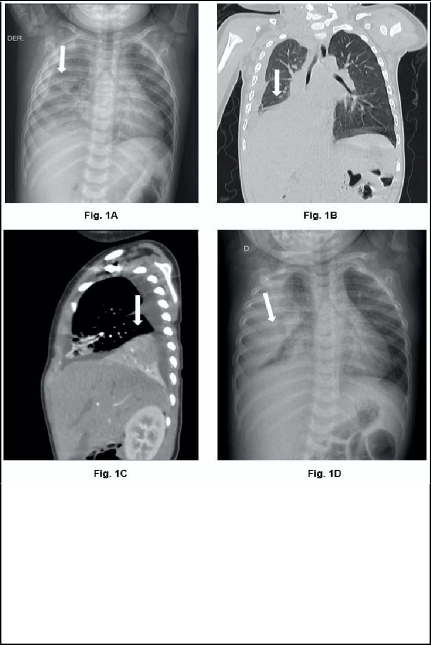
Figure 1.
A: AP chest X-ray. Diffuse linear pattern in the right lung parenchyma was observed.
B: Contrast-enhanced CT scan, coronal section. Consolidative image in the right lower lobe, with air bronchogram, heterogeneous post-contrast enhancement, and irregular intralesional hypodense areas.
C: Contrast-enhanced CT scan, sagittal plane. Ground-glass pattern in the middle lobe with homogeneous enhancement after contrast agent administration.
D: AP chest X-ray. Radiopacity of the right lung field with obliteration of the ipsilateral costophrenic angle.
cough and intermittent fever that partially subsided with antipyretics and antibiotics. Signs of respiratory distress such as tachypnoea, sub- costal retractions, and nasal flaring were added, and he was admitted to hospital.
The initial assessment us- ing the Wood-Downes score was 6 points, corresponding to moderate respiratory com- promise. He also presented with tachycardia and de- creased vesicular murmurs.
Tests for tuberculosis, including purified protein derivative (PPD) and poly- merase chain reaction (PCR), respiratory syncytial virus, mycoplasma, and SARS-CoV-2 (PCR), all
yielded negative results. Chest radiography revealed diffuse linear lesions in the right lung parenchyma, as- sociated with decreased lung volume and blurring of the right cardiophrenic and costodiaphragmatic angles (Fig. 1A).
Chest computed tomog- raphy with intravenous con- trast showed a condensative lesion with an air broncho-
gram in the right lower lobe with heterogeneous enhancement, presenting irregular hypodense areas suggestive of a necrotic process (Fig. 1B). In addition, a ground-glass pattern associated with thickening of the interlobular septa in the middle lobe with homogeneous enhancement (Fig. 1C), accompanied by moderate right pleural effusion with loculated characteristics.
Chachapoya-Rivas L.F., Chávez-Cruz N.E., Morales-Mayorga H., Serrano-Concha K.P., Acosta-Farina D.B. Organic foreign body as an unusual cause of complicated pneumonia in an infant: A case report.
Arch Med (Manizales). 2026;26(1)
Due to a poor response after 6 days of treatment with ceftriaxone and clindamycin, persistent fever spikes, and a progressive in- crease in supplemental oxygen, a chest X-ray was repeated, showing increased radiopacity and obliteration of the right costophrenic angle (Fig. 1D). Therefore, video-assisted thoracic surgery (VATS) was performed.
During CTAV, pleuroparietal and pleuro- visceral adhesions were observed, causing entrapment of the pulmonary lobes of the right hemithorax, pleural effusion in the process of organization, and 200 ml of yellowish fluid, which was collected for study and culture.
Complete pulmonary decortication was per- formed to release the trapped parenchyma. An organic foreign body was identified in the paren- chyma of the right lower lobe, directed towards the ipsilateral pleura (Fig. 2A). The foreign body was carefully removed using a trocar, confirming its nature as compatible with a grass awn (Fig. 2B). The postoperative period was monitored in an intermediate care unit.
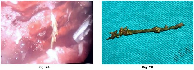
Figure 2.
A: Vegetable CE in the virtual space between the parietal and visceral pleura, with abundant pleural fluid supernatant on fibrin (CTAV).
B: Vegetable CE extracted (grass spikes).
In the immediate postoperative period, the patient presented with subcutaneous emphy- sema due to a bronchopleural fistula (Fig. 3A), which was managed with negative pressure
of 15 cmH2O in the pleural drain. Given the rapid progression to the posterior thorax and abdominal regions within 24 h (Fig. 3B), high- flow supplemental oxygen (FiO2 at 100%) was administered, and the patient was transferred to the intensive care unit due to the risk of de- compensation.
At 48 h, there was complete remission of em- physema (Fig. 3C), and supplemental oxygen was administered via a nasal cannula. Chest drainage was maintained for six days. He com- pleted a course of ceftriaxone and vancomycin with good clinical and respiratory mechanical evolution, allowing him to be transferred to a less complex area.
Due to the intraoperative findings, the family member was questioned again and reported that two months earlier, the child had expe- rienced transient asphyxia after probably in- gesting grass in the garden of his home; this resolved immediately after the maneuvers were performed, but a persistent cough remained, requiring several episodes of outpatient man- agement. The patient was discharged without complications and returned to the outpatient clinic one month later with good clinical and surgical progress to date (Fig. 3D).
Chachapoya-Rivas L.F., Chávez-Cruz N.E., Morales-Mayorga H., Serrano-Concha K.P., Acosta-Farina D.B. Organic foreign body as an unusual cause of complicated pneumonia in an infant: A case report.
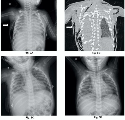
Figure 3.
A: AP chest X-ray. Right subcutaneous emphysema occurred 6 hours postoperatively.
B: Contrast-enhanced CT scan, coronal section. Subcutaneous emphysema in the right abdominal and thoracic walls extending to the posterior chest wall muscles [plain chest CT scan].
C: AP chest X-ray. Total remission of subcutaneous emphysema occurred at 48 h.
D: AP chest X-ray. Postoperative radiographic follow-up was performed in the outpatient clinic.
Arch Med (Manizales). 2026;26(1)
Bronchoaspiration of foreign bodies is one of the main pediatric respiratory emergencies in Ecuador [8]. Among these, plants such as grass awns behave differently owing to their morphol- ogy with barbs [9], which gives them migratory capacity and increases the risk of serious com- plications compared to inert objects [10].
Although uncommon, their potential to pass through bronchopulmonary structures to the pleura and chest wall has been described, similar to the present case [11]. Diagnosis is
often difficult owing to the non-specific clinical presentation and radiotransparency of plant bodies [12], which delays their detection by conventional radiography [13].
In the presence of respiratory symptoms that overlap with bacterial pneumonia and a lack of response to antibiotics, suspicion of a foreign body should be raised [14], as indicated in in- ternational protocols and paediatric emergency guidelines, which recommend considering this possibility in cases of persistent or poorly evolv- ing pneumonia [15].
Chachapoya-Rivas L.F., Chávez-Cruz N.E., Morales-Mayorga H., Serrano-Concha K.P., Acosta-Farina D.B. Organic foreign body as an unusual cause of complicated pneumonia in an infant: A case report.
Arch Med (Manizales). 2026;26(1)
The persistence of respiratory symptoms that do not respond to antibiotics should raise suspi- cion of a foreign body [16], particularly in cases of prolonged or poorly evolving pneumonia [17]. These represent the main complications increase hospital stays, antibiotic use, and the need for surgery [15,18].
Pulmonary entrapment due to pleurovisceral adhesions observed in CTAV is consistent with the mechanisms described for prolonged bron- chial obstructions, with retained secretions and secondary infection progressing to empyema and pleural fibrosis [9].
The intraoperative findings of the plant body confirmed its migratory behavior, favored by the unidirectional structure of the spikes, which are capable of penetrating the parenchyma and pleura, causing erosion and major complica- tions [12].
Tension pneumothorax due to persistent air leakage has also been described [18]. In this case, postoperative subcutaneous emphysema occurred, a complication documented after the surgical removal of migrated bodies [14].
The intraoperative findings of the plant body confirmed its migratory behavior, favored by the unidirectional structure of the spikes, which
are capable of penetrating the parenchyma and pleura, causing erosion and major complica- tions [12].
Tension pneumothorax due to persistent air leakage has also been described [18]. In this case, postoperative subcutaneous emphysema occurred, a complication documented after the surgical removal of migrated bodies [14].
CE aspiration can present as persistent pneumonia that is difficult to treat. Its location between the lung parenchyma and pleural space, which is rarely documented in the liter- ature, emphasizes the need for a high index of suspicion. Video-assisted thoracoscopy is an effective and minimally invasive diagnostic and therapeutic method.
Consent for publication: Informed consent was obtained from the patient.
Conflict of interest: The authors declare no conflict of interest.
Source of funding: No.
Author contributions: The authors contrib- uted to and approved the final version of this manuscript.
Wang Q, Kong X, Wang G, Dai J, Li Y, Wu C, et al. A real-world study of foreign body aspiration in children with 4227 cases in Western China. Sci Rep. 2024;14(1). https://doi.org//10.1038/s41598-024-65876-7
Zhu Y, Fan Q, Cheng L, Chen B. Diagnostic Errors in Initial Misdiagnosis of Foreign Body Aspiration in Children: A Retrospective Observational Study in a Tertiary Care Hospital in China. Front Pediatr. 2021;15;9. https://doi.org//10.3389/fped.2021.694211
Rance A, Mittaine M, Michelet M, Martin Blondel A, Labouret G. Delayed diagnosis of foreign body aspiration in children. Arch Pediatr. 2022;29(6):424-428. https://doi.org//10.1016/j.arcped.2022.05.006
Cai H, Gao J. Clinical analysis of special types of tracheobronchial foreign bodies in children. Front Pediatr. 2024;12. https://doi.org/10.3389/fped.2024.1395629
Louis M, Tapia R, Grabill N, Bongu N, Singh H, Hastings JC. Missed Foreign Body Aspiration: Fentanyl Patch Leading to Severe Pneumonia and Empyema. Cureus. 2025;17(1):e77931. https://doi.org//10.7759/cureus.77931
Razafimanjato NNM, Ralaivao RA, Ravelomihary TDN, Hunald FA, Rakotovao JLH. Pneumonectomy in a child due to belated diagnosis of foreign body aspiration: a case report. J Med Case Rep. 2021;15(1). https://doi.org//10.1186/s13256-021-03015-w
Chachapoya-Rivas L.F., Chávez-Cruz N.E., Morales-Mayorga H., Serrano-Concha K.P., Acosta-Farina D.B. Organic foreign body as an unusual cause of complicated pneumonia in an infant: A case report.
Arch Med (Manizales). 2026;26(1)
Richard N, Paygambar A, Ducou Le Pointe H, Biaz S, Corvol H. Hordeum murinum aspiration revealed by a pneumopleurocutaneous fistula in a 15-month-old infant. BMC Pediatr. 2021;21(1). https://doi.org//10.1186/s12887-021-03016-0
Córdova Neira F, Dávila Tapia B: Ingestión de cuerpos extraños en niños. Rev Ateneo. Vol 24, No.(1) pág. 157-168.
Szabó H, Kálmán A. Migration of an aspirated grass inflorescence through the chest wall in a 3-year-old boy. Pediatr Pulmonol. 2021;56:344-6 https://doi.org//10.1002/ppul.25178
Newson TP, Parshuram CS, Berkowitz RG, Auldist AW, Robinson PJ. Tension pneumothorax secondary to grass head aspiration. Pediatr Emerg Care. 1998;14(4):287-9. https://doi.org//10.1097/00006565-199808000-00013
Asadi Gharabaghi M, Asadi Gharabaghi M, Firoozbakhsh S. Empyema caused by foreign body aspiration. BMJ Case Rep. 2012;2012. https://doi.org//10.1136/bcr-2012-007760
Chen Y, Li L, Wang C, Zhang Y, Zhou Y. Necrotizing Pneumonia in Children: Early Recognition and Management. 2023;12(6):2256.: https://doi.org//10.3390/jcm12062256
Vlahova A, Antonova Z, Rangelov E, Kartulev N, Oparanova V, Gabrovska N, et al. Surgical Treatment of Lung Abscess Due to an Awn Aspiration in a 9-Year-Old Child: A Case Report. Children. 2023;10(6):910. https://doi.org/10.3390/children10060910
Joseph L, Assous M, Dreifuss K, Goldberg S, Picard E. Complicated Pediatric Pneumonia With Eikenella Caused by Foreign Body Aspiration: Institutional and Literature Review. Case Rep Infect Dis. 2025;2857930. https://doi.org//10.1155/crdi/2857930
Lobeiras Tuñón A. Ingesta-aspiración de cuerpo extraño. En: Protocolos diagnósticos y terapéuticos en ur- gencias de pediatría. 4ª ed. Madrid: Sociedad Española de Urgencias de Pediatría (SEUP); 2024. p. 1-12. https://seup.org/wp-content/uploads/2024/04/26_Ingesta_CE_4ed.pdf
Martinez Diaz M, Ibañez Pradas V, Couselo Jerez M, Valdés Diéguez E. Airway exploration when suspected foreign body aspiration. Is it always indicated? Journal of Pediatric Surgery Open. 2024;8:100163. https://doi.org//10.1016/j.yjpso.2024.100163
Mahmood U, Talat N, Ur Rehman W, Khalid R. “Innovative multidisciplinary care in pediatric magnet aspiration: A case report.” Int J Surg Case Rep. 2025;126. https://doi.org//10.1016/j.ijscr.2024.110814
Mîndru DE, Păduraru G, Rusu CD, Țarcă E, Azoicăi AN, Roșu ST, et al. Foreign Body Aspiration in Children— Retrospective Study and Management Novelties. Medicina (Lithuania). 2023;59(6). https://doi.org//10.3390/medicina59061113
Yankov G, Alexieva M, Valev D, Mekov E. Development of Organized Pleural Empyema as a Result of Occult Foreign Body Aspiration. Folia Med (Plovdiv). 2023;65(6):1000–4. https://doi.org//10.3897/folmed.65.e91076
Bjerregaard AT, Holm JK, Clausen NG. Time From Suspected Foreign Body Aspiration to Bronchoscopy at Odense University Hospital During a 5-Year Period. Acta Anaesthesiol Scand. 2025;69(6). https://doi.org//10.1111/aas.70053
Markov S, Markova P, Karavelikova I, Halacheva H. Foreign Bodies in Lower Airway in Children: Brief Review and Clinical Experience. Children. 2025; 12(1):67. https://doi.org//10.3390/children12010067
Shilpakar R, Shrestha N, Shrestha D. Bronchial foreign body presenting as pneumothorax and pneumomediasti- num in a young child. Clin Case Rep. 2024;12(11). https://doi.org//10.1002/ccr3.9505